
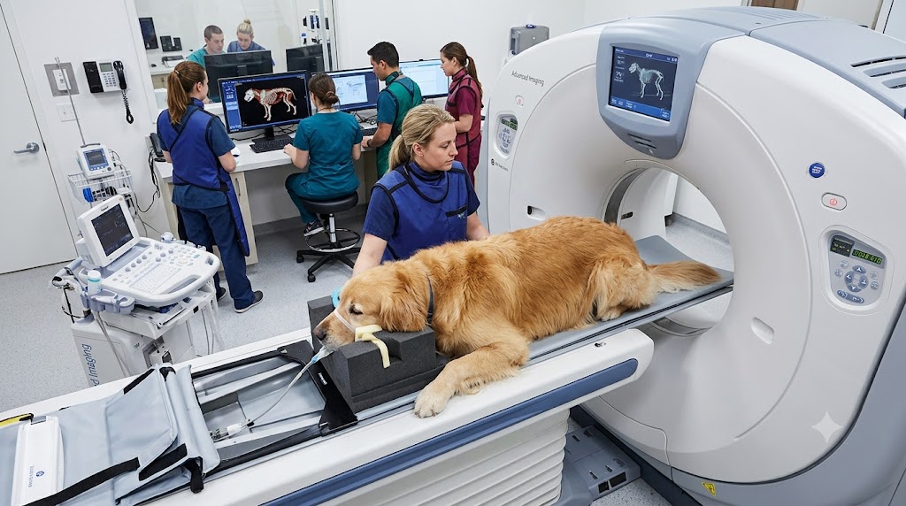
The Region’s Most Advanced Veterinary CT Technology
Advanced diagnostic imaging is coming closer to home.
West Texas Veterinary Specialists is bringing 64-slice helical CT with 128-slice reconstruction to veterinary patients throughout West Texas and the surrounding region, providing exceptional speed, image quality, and diagnostic detail for complex medical and surgical conditions.
This advanced technology allows detailed cross-sectional evaluation of anatomy that may be difficult to assess with conventional imaging and can help specialists more precisely characterize disease, plan procedures, and determine appropriate next steps in care.
Our referral-only model allows CT findings to become part of a coordinated diagnostic plan developed in collaboration with your primary veterinarian and specialty team.
What is a CT Scan?
Computed tomography, or CT, uses X-rays and advanced computer processing to create detailed cross-sectional images of the body.
Unlike a traditional radiograph, which produces a two-dimensional image in which structures may overlap, CT generates thin image slices that allow specific areas to be evaluated in much greater detail. Images can also be reconstructed in three-dimensional (3D) and multiple planes when useful for diagnosis or procedural planning.
Advanced CT Technology for West Texas
Our 64-slice helical CT scanner with 128-slice reconstruction represents a major advancement in veterinary diagnostic imaging for the region.
Rapid image acquisition and high-resolution reconstruction allow detailed evaluation of complex anatomy, including small or difficult-to-visualize structures. This can help define the location and extent of disease and provide detailed information for diagnostic, surgical, oncologic, and minimally invasive procedural planning.
The combination of advanced imaging technology and specialty expertise allows CT findings to be evaluated not simply as images, but as part of the patient’s overall medical or surgical condition.
When Is CT Recommended?
CT can provide valuable information for a wide range of medical and surgical conditions, including:
Head & Airway
Chronic nasal disease, nasal masses, skull and facial abnormalities, middle and inner ear disease, and airway disorders.
Chest & Abdomen
Pulmonary nodules and masses, thoracic abnormalities, abdominal masses, vascular abnormalities, and selected organ and soft-tissue disorders.
Orthopedic & Neurologic
Complex fractures, bone lesions, joint abnormalities, spinal disease, and surgical planning.
Cancer Evaluation
Characterization of masses, evaluation of disease extent, selected metastatic screening, and treatment or surgical planning.
The most appropriate imaging study depends on the patient’s condition. Your veterinarian and our specialty team can help determine whether CT or another diagnostic modality is best suited to answer the clinical question.
CT as Part of a Complete Diagnostic Plan
Advanced technology can provide remarkable images, but the value of CT extends beyond producing those images.
CT findings must be interpreted within the context of the patient’s clinical history, physical examination, laboratory results, and other diagnostic findings to determine their clinical significance and how they should influence the next steps in care.
When appropriate, CT can also be integrated with our other specialty and diagnostic services, including Internal Medicine, Surgery, Oncology, Ultrasound & Echocardiography, Endoscopy, and Fluoroscopy.
What to Expect
Most veterinary patients require sedation or general anesthesia for CT because remaining completely still is essential for obtaining diagnostic-quality images.
Depending on the condition being evaluated, intravenous contrast may also be administered to improve visualization of blood vessels, organs, masses, and other soft-tissue structures.
The scan itself is typically completed quickly, often within seconds to a few minutes, although preparation, sedation or anesthesia, contrast administration when indicated, and recovery mean your pet will remain with us longer than the actual scan time.
Our team will provide specific preparation and fasting instructions before your pet’s appointment.
Our Collaborative Approach
West Texas Veterinary Specialists is a referral-only outpatient specialty center. Patients may be referred specifically for CT imaging, or CT may be recommended as part of a broader specialty evaluation.
Imaging findings and recommendations are communicated to the referring veterinarian so results can be incorporated into the patient’s ongoing diagnostic and treatment plan.
FREQUENTLY ASKED QUESTIONS
Why might my pet need a CT scan?
CT may be recommended when more detailed imaging is needed than can be obtained with traditional radiographs or when the location or nature of an abnormality is particularly well suited to cross-sectional imaging.
It is commonly used to evaluate conditions involving the head, nasal passages, chest, bones, joints, spine, masses, and other complex anatomy.
What is the difference between CT and a regular X-ray?
Traditional radiographs produce two-dimensional images in which anatomical structures may overlap. CT creates detailed cross-sectional images that allow structures to be evaluated individually and reconstructed in multiple planes.
What is special about the CT technology at West Texas Veterinary Specialists?
Our center is equipped with a 64-slice helical CT scanner with 128-slice reconstruction, bringing some of the most advanced veterinary CT capabilities available to West Texas and the surrounding region.
Its rapid image acquisition and high-resolution reconstruction provide exceptional anatomic detail that can be valuable for complex diagnostic evaluations and surgical or procedural planning.
Will my pet need anesthesia?
Most veterinary patients require sedation or general anesthesia to remain completely still during CT imaging. The appropriate protocol depends on your pet’s condition and the type of study being performed.
Will my pet receive contrast?
Some CT studies require intravenous contrast to improve visualization of blood vessels, organs, masses, and other soft tissues. Whether contrast is recommended depends on the area being evaluated and the clinical question being investigated.
How long will my pet’s CT appointment take?
The CT scan itself can often be completed quickly, but preparation, sedation or anesthesia, contrast administration when indicated, and recovery add additional time. Our team will provide an anticipated schedule based on the type of study your pet requires.
Can my pet go home the same day?
Most patients undergoing CT at West Texas Veterinary Specialists return home the same day after appropriate recovery from sedation or anesthesia.
We are an outpatient specialty center and do not provide overnight hospitalization or intensive care.
How will I receive the results?
CT findings and recommendations are communicated to your referring veterinarian and incorporated into your pet’s diagnostic and treatment plan. The timing of final results may depend on the type and complexity of the study.
Ready to Refer a Patient?
If your veterinarian has recommended CT or advanced diagnostic imaging, our team is here to help coordinate your pet’s referral.