
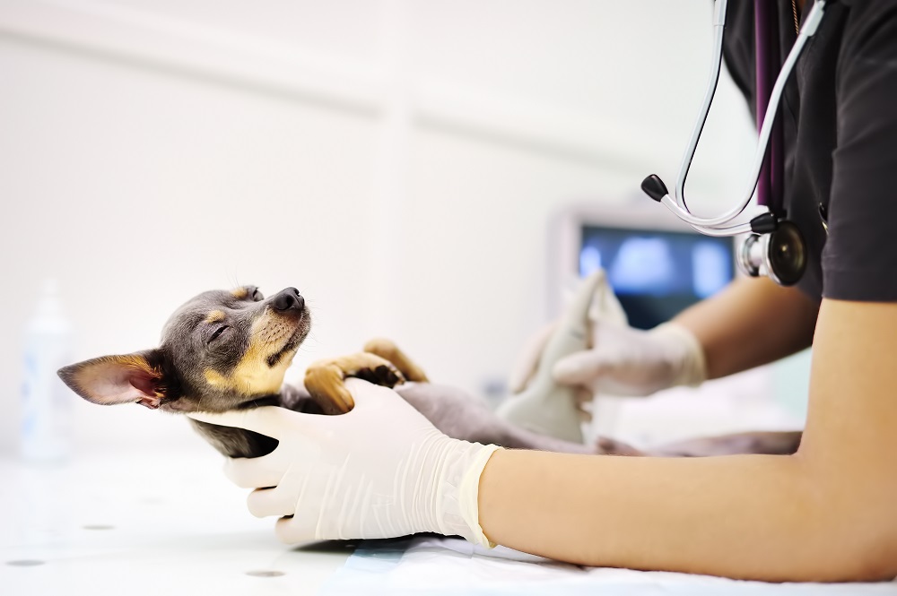
Specialist-Performed Imaging. Whole-Patient Interpretation.
Ultrasound can reveal remarkable detail about a pet’s internal organs and cardiovascular system—but obtaining an image is only part of the diagnostic process.
At West Texas Veterinary Specialists, abdominal ultrasound and echocardiography are performed and interpreted by Kirk Sears, DVM, DACVIM (SAIM), a board-certified veterinary internal medicine specialist with extensive experience in diagnostic ultrasonography.
This allows imaging findings to be evaluated alongside the patient’s history, physical examination, laboratory abnormalities, medications, and other diagnostic results—helping determine not only what an abnormality is, but whether it is clinically significant and what should happen next.
Why Specialist-performed Ultrasound Matters
Ultrasound has become increasingly available in veterinary medicine, but the diagnostic value of an examination depends on more than the technology itself—it also depends on the training and experience of the person performing and interpreting the study.
Ultrasound is a dynamic, real-time examination. The person performing the study determines which structures to examine more closely, which measurements to obtain, how findings relate to one another, and whether additional views or diagnostic samples are needed as the examination progresses.
As a board-certified veterinary internist, Kirk Sears, DVM, DACVIM (SAIM), combines advanced specialty training in complex medical disease with extensive experience performing and interpreting ultrasound examinations.
This whole-patient approach helps distinguish clinically meaningful abnormalities from incidental findings, guides additional diagnostic testing, and determines appropriate next steps in care.
Abdominal Ultrasound
Abdominal ultrasound provides detailed, real-time evaluation of the liver and gallbladder, spleen, kidneys and adrenal glands, gastrointestinal tract, pancreas, abdominal lymph nodes, urinary tract, reproductive organs, blood vessels, and surrounding tissues without the use of ionizing radiation.
Ultrasound can help identify changes in organ structure, masses or nodules, enlarged lymph nodes, abnormal fluid accumulation, gastrointestinal abnormalities, and other changes that may not be apparent with traditional radiographs.
Ultrasound-Guided Diagnostic Sampling
One of the important advantages of real-time ultrasound is the ability to precisely guide diagnostic sampling when appropriate.
Based on the ultrasound findings and the patient’s condition, Kirk Sears, DVM, DACVIM (SAIM), may recommend ultrasound-guided fine-needle aspiration, tissue biopsy, cystocentesis, or sampling of abnormal fluid.
Real-time ultrasound guidance allows specific organs, masses, lymph nodes, or other abnormalities to be targeted while avoiding surrounding structures. Sampling is recommended when it is expected to provide clinically useful information and help guide diagnosis or treatment.
Echocardiography
Echocardiography is a specialized ultrasound examination of the heart that provides real-time information about cardiac structure and function.
An echocardiogram allows evaluation of the heart chambers, valves, cardiac muscle, blood flow, and surrounding structures and can help identify or characterize many forms of heart disease.
Echocardiography may be recommended for patients with:
- A heart murmur or abnormal heart sounds
- Suspected or known heart disease
- Cardiac enlargement on radiographs
- Abnormal heart rhythm
- Unexplained respiratory signs
- Fluid accumulation that may be related to heart disease
- Certain breed-associated cardiac concerns
At West Texas Veterinary Specialist, echocardiographic findings are interpreted within the context of the patient’s complete medical picture, helping determine the significance of cardiac abnormalities and guide recommendations for additional testing, treatment, or monitoring when appropriate. Further, while Dr. Sears is comfortable interpreting the vast majority of cardiac examinations, he occasionally submits studies to a board-certified cardiologist for further interpretation.
What To Expect
Most abdominal ultrasound and echocardiographic examinations are non-invasive and can be performed without general anesthesia.
A specific area of fur is typically clipped to allow appropriate contact between the ultrasound probe and the skin. Depending on the patient’s temperament, medical condition, and whether diagnostic sampling is needed, mild sedation may occasionally be recommended.
If ultrasound-guided aspiration or biopsy is appropriate, our team will discuss the procedure and any additional preparation or sedation that may be required.
Our Collaborative Approach
West Texas Veterinary Specialist is a referral-only outpatient specialty center.
Patients may be referred specifically for an abdominal ultrasound or echocardiogram, or imaging may be recommended as part of a broader Internal Medicine evaluation.
Findings, diagnostic results, and recommendations are communicated to the referring veterinarian so they can be incorporated into the patient’s ongoing diagnostic and treatment plan.
Our role is to provide specialty expertise while maintaining the important relationship and continuity of care between pets and their primary veterinary team.
FREQUENTLY ASKED QUESTIONS
Please use an accordion/expand-and-collapse format for this section.
Why might my pet need an abdominal ultrasound?
Ultrasound may be recommended to investigate abnormal laboratory results, chronic gastrointestinal signs, weight loss, abdominal pain, labwork changes involving the liver or kidneys, suspected masses, abnormal fluid accumulation, or other conditions affecting the abdominal organs.
What is the difference between ultrasound and an X-ray?
Radiographs and ultrasound provide different types of information. Radiographs are particularly useful for evaluating bones, the lungs, and the overall size and position of structures, while ultrasound provides detailed real-time evaluation of many internal organs and soft tissues.
The most appropriate imaging test depends on the clinical question being investigated.
Why does it matter who performs my pet’s ultrasound?
Ultrasound is a dynamic examination rather than simply a collection of static images. The person performing the study makes real-time decisions about which structures require closer evaluation, what measurements or additional views are needed, and whether diagnostic sampling should be considered.
At West Texas Veterinary Specialist, examinations are performed and interpreted by Kirk Sears, DVM, DACVIM (SAIM), allowing imaging findings to be instantly interpreted and integrated with his advanced knowledge in complex medical disease and the patient’s overall clinical picture.
What is the difference between an abdominal ultrasound and an echocardiogram?
Both use ultrasound technology, but they evaluate different areas of the body. Abdominal ultrasound examines organs within the abdomen, while echocardiography is specifically designed to evaluate the structure and function of the heart.
Will my pet need sedation?
Most abdominal ultrasounds and echocardiograms can be performed without sedation. Mild sedation may be recommended in some cases depending on the patient’s temperament, fear/anxiety level, medical condition, or whether diagnostic sampling is planned. Sedation is a very safe procedure that decreases anxiety to the patient and improves image study quality.
Can you take samples during the ultrasound?
Yes. When appropriate, ultrasound can be used to guide fine-needle aspiration, tissue biopsy, urine collection, and sampling of abnormal fluid.
Whether sampling is recommended depends on the ultrasound findings and the individual patient’s condition. Ultrasound-guided sampling requires training and skill, and it will only be recommended when the potential diagnostic or therapeutic benefit of the procedure significantly outweighs the risk.
How will I receive the results?
Findings and recommendations are communicated to the referring veterinarian so they can be incorporated into your pet’s ongoing diagnostic and treatment plan. In addition, you can schedule an in-person consultation with the specialist once the procedure has concluded. Our team will also discuss the expected process for results based on the type of referral and services performed.
Ready To Refer A Patient?
If your veterinarian has recommended abdominal ultrasound, echocardiography, or other advanced diagnostic procedure, our team is here to help coordinate your pet’s referral.